Dissections Overview
Throughout all the dissections we did, all of learned a lot about the anatomy of different animals, and how they must adapt, both externally and internally, to survive in the environment they live in. We saw these changes as we progressed through the fish, then frog, and finally the rat and pig. Below, we will show the documentation of each dissection (through pictures and video), and additional information about what we learned when dissecting. We will show this information in the order of which we did the dissections.
The Fish Dissection
|
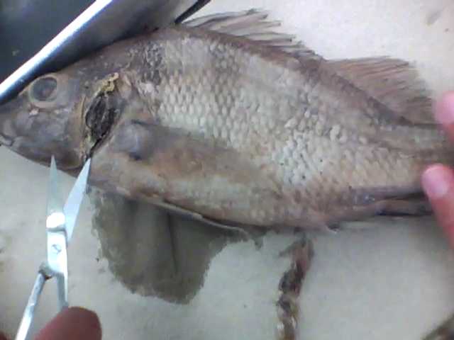
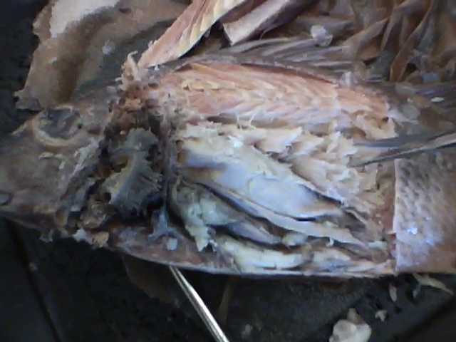
Shown on both sides of this text, are pictures of the fish before, and after we opened it up. On the right, we had already begun to remove the operculum, and on the left, we had finished removing all the skin and flesh covering the organ systems. Because of the tough scales, opening the fish proved a bit challenging for us, and resulted in a messier opening.
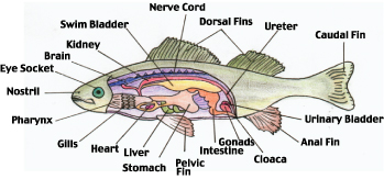
Included under our pictures, are labeled diagrams of the fishes external (left) and internal (right) systems. |
|
|
|
Dissection VideosThe video directly left, titled "IMG 0004" shows Frank, Doug, and I opening up the fish. We are making the initial incisions, as directed by the handout we received.
To the left, the video titled "IMG 0008" shows us making a crude attempt to identify organs after removing the skin of the fish. We have some trouble identifying organs, and initially mislabel the swim bladder as the stomach. Later (not shown), we realize our error, and label everything correctly. This being the first dissection, we did not realize the importance of the hand out, and how it showed us all the organs. |
The Frog Dissection
Smoother Than the Last
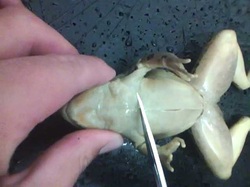
Overall, the dissection of the frog was much cleaner, and went more smoothly that that of the perch. We paid closer attention to the instructions on the pamphlet, and were able to open the frog quickly. On top of that, we were able to extract all of the organs, almost completely in-tact. This time, we knew what we were doing (at least more than we did with the fish). We probably could have done a better job, but our work was fantastic in comparison to the perch.
|
|
|
Diagram of the Frog's Internals
As shown by the diagram in comparison to the videos aboce, we were able to keep all the frog's organs intact for extraction, unlike the fish. Although we were a bit brutal in opening the chest cavity, we did it delicately enough to be able to take out the organs under it in one piece.
|
The internal organs of the frog |
The Rat Dissection
First One
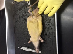
Being the first mammal, and strictly land animal we dissected, the rat was a fun dissection. We saw the similarities to between how our organs look, and theirs. The rat's body was adapted for land only, and this was shown by its shape. The rat was built for running (or scurrying along on its paws), and has sharp nails on its feet for climbing. Also, it has a tail for balance. We noted how this was a change from the frog, who had webbed feet, and smooth skin for swimming.
|
|
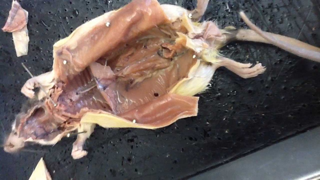
Dissecting the rat went flawlessly; After having done two previous dissections the rat seemed easy. This was because of how big the rat was, and because cutting through it was not difficult (the rats skin and flesh were soft). We did this dissection quickly, and without error.
This can be seen by the clean body cavity of the rat, and how in-tact all the organs are. |
|
|
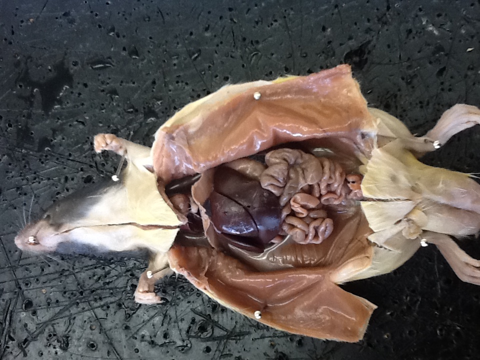
On the left we have a picture of the rat, opened up. You can see all of its organs clearly, from the diaphragm to the anus.
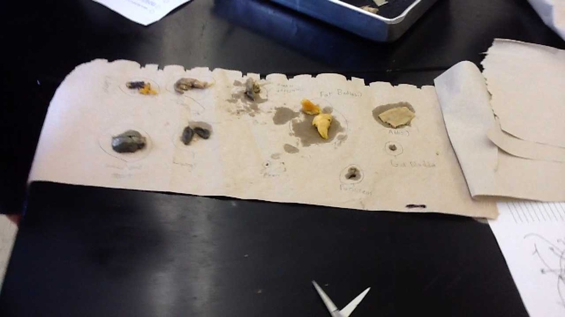
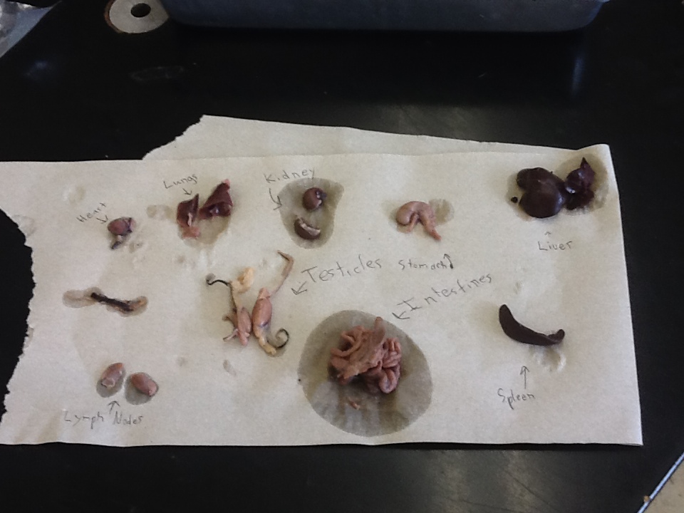
On the right, we have all the organs laid out, labeled, after we removed them from the rat. Next to them, is the body cavity of the rat, which has been emptied of all organs. |
|
|
The Fetal Pig
The Final Dissection
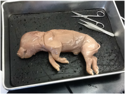
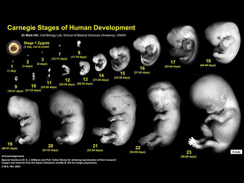
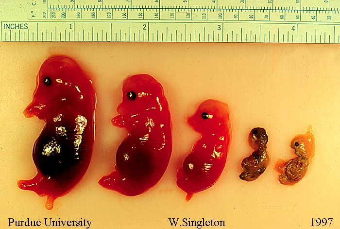
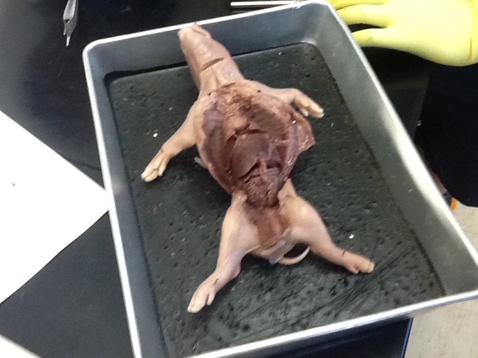
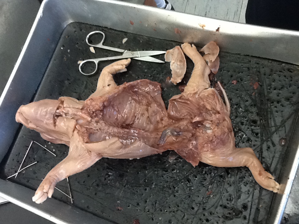
Dissecting the fetal pig was interesting, not only because it was the largest of animals we had used, but because it was at a stage where you could see it taking distinct shape as a pig. It was no longer at a stage that it looked similar to us, or birds, or anything else. We determined, through online research, that the pig we dissected was between 86 and 100 days old. While dissecting, we had brief pangs of guilt, because the pig looked so innocent, and babyish. However, we got over this, and proceeded. This dissection also went without any hiccups, because by the time we dissected the pig, we knew how to do it right.
|
An interesting thought we kept in mind while dissecting was the similarity that this pig would have had to a human fetus only a few weeks before this dissection. As a group, we discussed briefly what it could mean about the origin of our species. When researching, we found pictures demonstrating the similarities and were very amazed. The human fetus is the picture on the left, and the pig is on the right.
|
|
|
|
|
Above, we have pictures and a video of the process of dissecting the pig.
Final Notes
As a group we would like to thank you, Mr. Devera, for allowing us to do these dissections. They were a fun project, and a fantastic way to end the year.
Sincerely,
Theo Madura, Frank Musso, and Doug Brown
Sincerely,
Theo Madura, Frank Musso, and Doug Brown
